Dental disease is among the most common illnesses seen in cats and dogs. Many pets suffering from dental disease show no apparent signs there is a problem. Our case this week outlines one such case of a hidden but serious dental issue in a dog.
During a routine annual examination in an otherwise healthy Golden Retriever, I noted an abnormality with one of the major chewing teeth: the upper 4th premolar (also called the upper carnassial) on the right side. This tooth had a fracture line across the length of the tooth. When probed, the fractured segment was confirmed to be completely separate from the rest of the tooth.
This type a fracture, called a slab fracture, is unique to this particular tooth. It occurs when the patient has chewed down on something firm and non-pliable, such as a bone, antler, or rock.
Deep slab fractures allow oral bacteria access to the central part of the tooth known as the pulp cavity. This is where the nerves and blood supply for the tooth are found. When bacteria enter this cavity, the pulp tissue reacts to the bacteria, and inflammation (swelling) of the pulp results. Because the pulp is trapped within the confines of the tooth, the swelling leads to pressure within the tooth, compromising the blood supply and ultimately causing a loss of vitality of the tooth.
When this large tooth loses vitality, the bacteria from the mouth can invade deeper and may exit the tooth through tiny openings at the tips of the roots, deep within the upper jaw. This can then lead to a tooth root abscess – a large, painful swelling filled with bacteria and pus that appears on the side of an affected pet’s face, just under the eye.
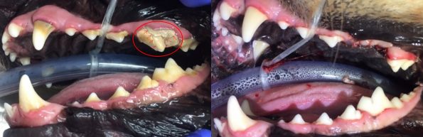
To address the fractured tooth, and prevent formation of a tooth root abscess, I recommended extraction of the affected tooth. The image above (left) shows the mouth before the circled tooth was extracted. On the right, the mouth is shown after extraction of the tooth. The remaining teeth have also been professionally scaled and polished.
After removing the tooth, it was clear to see the pulp tissue was compromised: it was brown in colour instead of the typical pink we see with healthy tooth pulp. The image below shows the tooth after extraction, pieced back together with 2 of the 3 roots shown (left), and the cut edge exposed, showing the pulp cavity (right) – the fragmented portion and third root are also shown.

Our patient is expected to recover uneventfully from the extraction, and since the tooth is fully extracted, there is no risk that a tooth root abscess will form. After the extraction site has healed, this patient will still be able to eat regular kibbles without difficulty.
To prevent slab fractures, do not allow your dog to chew on hard objects. As a rule of thumb, if an object cannot be flexed or bent with your hands, it is probably too hard for your dog’s teeth!