Today we are reviewing the case of a 7 year old female cat that presented to our clinic with rapid breathing. Upon examination of this sweet cat, I counted the respiration rate at 60 breaths per minute – approximately double the maximum normal respiration rate.
When listening to this patient’s chest, I noted wheezing in the lungs in the upper parts of the chest and minimal to no noise in the lower parts of the chest. The heart was quiet and difficult to hear.
As is typically recommended in any case of breathing changes, x-rays of the chest were performed. The x-rays revealed a condition I was worried I might find: pleural effusion. Pleural effusion is fluid (effusion) in the chest cavity (pleural cavity), or the space between the chest wall and the lungs. Normally the lungs fill the entire chest cavity and there should not be an accumulation of fluid in this area. With the fluid present in the chest, it is harder to hear the heart and lungs during examinations.
Pleural effusion can occur from various conditions including heart failure, protein deficiencies, and tumors in the chest. This is a potentially life-threatening condition because the fluid takes up the space the lungs need to expand as they fill with air. It becomes difficult for the body to get enough oxygen from the reduced volume of air in the lungs.
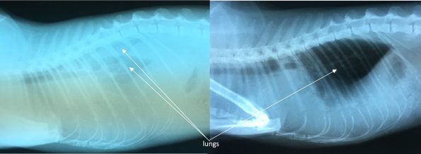
In this case, it was difficult to determine from the intial x-ray whether the fluid was related to a heart problem. This is because the fluid on the x-ray blends in with the heart and prevents veterinarians from assessing the size and shape of the heart and blood vessels. Therefore, I performed a procedure called a thoracocentesis. This procedure involves inserting a needle through the chest wall and drawing off the excessive fluid. See the image for the x ray before this procedure (left) and after (right).
After draining over 200mls of fluid, our patient was feeling better and more energetic because her lungs could expand more freely. Repeating the radiographs showed the heart was enlarged. Based on this and other characteristic changes in the chest, I concluded this patient was in heart failure.
Heart failure means that the heart is not efficiently pumping the blood forward in the circulatory system. As a result, blood builds up in the veins and excess fluid begins to leak out of the circulation into the tissues or chambers in the body. In the case of our patient, the fluid appeared in the chest cavity. In other cases of heart failure, we may see excessive fluid in the lungs themselves or the abdominal cavity.
To treat the heart failure, I prescribed a diuretic – a medication that works on the kidneys to increase fluid release from the blood into the urine. I also prescribed a medication that helps to reduce the resistance in the arteries to ease the workload on the heart.
After two weeks on the medication, I repeated the x-rays and was pleased to see the pleural fluid had not returned.
This patient is now feeling better, gaining weight back, and has more energy with the heart failure treatment. Because there is no cure for heart failure, the medical treatment will need to continue life long. Thankfully, once medicated appropriately, many pets continue to enjoy a high quality of life for a long time while in dealing with heart failure!